Thursday, 13 September 2012

DISEASES & PATHOGEN

Diseases

A disease can modify or affect chemical reactions in the body and set it to disequilibrium

Non-communicable: self-inflicted or created by environmental conditions, such as dietary deficiency diseases or cancers

E.g:
  1. Scurvy
  2. Rickets
  3. Obesity
  4. Anorexia
  5. Coronary heart diseases

Communicable: Due to agents such as viruses or bacteria, which are passed between organisms

E.g:
  1. Cough
  2. Flu
  3. Malaria
  4. Athlete's Foot

Pathogen 

cause the disruption of the equilibrium of the body & the immune system helps to restore & maintain the equilibrium
** Not all bacteria are pathogen! (yaklut) But pathogen can be bacteria** 

  • Organisms that cause disease and lives in or on another organism.
  • When they infect an organism, they
  1. Destroy cells
  2. Produce toxins
  3. Interfere with normal functions in organism
  4. Invokes immune response that, if excessive, harms the organism.
  • To infect an organism, the organism must 
  1. able to survive the transmission from one host to another. 
  2. able to enter the host. 
  3. virulent – the ability to overcome body defenses (through its own replication) & only need to be initially present in small numbers. 

Different classes of pathogens:

  • Bacteria 
  • Viruses 
  • Protozoa 
  • Parasites
  • Fungi 


Further Readings:
Botox - http://dermatology.about.com/od/cosmeticprocedure/a/botox.htm
HIV/ AIDS - http://www.google.com.sg/url?sa=t&rct=j&q=&esrc=s&source=web&cd=3&sqi=2&ved=0CEQQFjAC&url=http%3A%2F%2Fen.wikipedia.org%2Fwiki%2FHIV%2FAIDS&ei=lnxSUNrSJYzorQeQ3YG4Bg&usg=AFQjCNF0DBD0fizNfrRaYD9L4NMDXgr8wg&sig2=udJp1yPcZJ9U5ZR0EyfePA

Monday, 3 September 2012

CLARIFYING QNS:)

1) Does diffusion occur instantly?

Diffusion - matter are made up of particles that are always moving. Thus, answer is no.

2) Difference between ingestion & digestion?

Ingestion - process of "eating" of taking in nutrients.
Digestion - mechanical, chemical

*similarity: occur at the same time.

3) What does it mean for intestine to be convoluted

** increase surface area to volume ratio

4) What is the difference between bile & bile salts?

In our level, we can use both. Bile salts is in bile:)

5) Function of semi-lunar valve and tricuspid valve?

Tricuspid Valve (Right Hand Side) --> preventing the back-flow of blood from the right ventricle to the right atrium of the heart

6) Heavy smoking affect heart?

nicotine--> hardening of vessels
cholestrol gets trap in the vessels.

Tuesday, 28 August 2012

TRANSPORT IN MAMMALS


Objectives:

  • Know that blood is pumped around the circulatory system by the action of the heart.
  •  Know that the heart is a muscular organ which requires its own supply of oxygen and nutrients (via coronary arteries)
  •  Label and identify the principle structures of a mammalian heart, including the valves within it and the inter-connecting blood vessels & associated valves.
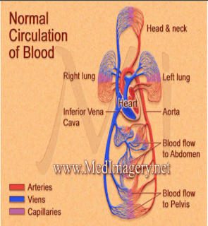
  •  Trace the path of blood through the circulatory system, in particular the pathway of oxygenated and deoxygenated blood through the heart.


HEART:


  •  Involuntary Muscle
  • Has 4 chambers
– Right & left atrium (single atrium/auricle)
– Right & left ventricles


Why is the left ventricle wall so thick?

  • The ventricle pumps blood at high pressure out to the arteries (to the lungs or other parts of the body)
  •  The pressure generated by the left ventricle is greater than that generated by the right ventricle as the systemic circuit is more extensive than the pulmonary circuit


Why is the muscle around the atria thinner than the muscle around the ventricles?

  • The ventricle pumps blood at low pressure out to the arteries (to the lungs or other parts of the body)
  •  The pressure generated by the left ventricle is lower thanthat generated by the rightventricle as the systemiccircuit is more extensive than the pulmonary circuit 


The Valve: 


  •  Ensure that the blood flows in the right direction 
  •   Very important; without them, the flow of blood would be chaotic 
  • Found in the heart and veins


Tricuspid Valve: 


  • Separates the right atrium from the right ventricle.
  • It opens to allow the deoxygenated blood collected in the right atrium to flow into the right ventricle.
  • It closes as the right ventricle contracts, preventing blood from returning to the right atrium; thereby forcing it to exit through the pulmonary valve into the pulmonary artery. 


BICUSPID VALVE(MITRAL VALVE)
  • Separates the left atrium from the left ventricle.
  • It opens to allow the oxygenated blood collected in the left atrium to flow into the left ventricle.
  • It closed as the left ventricle contracts, thereby forcing it to exit through the aortic valve into the aorta.


PULMONARY VALVE
  • Separates the right ventricle from the pulmonary artery.
  • As the ventricles contract, it opens to allow the de-oxygenated blod collected in the right ventricle to flow to the lungs. 
  • It closes as the ventricles relax, preventing blood from returning to the heart.


SUPERIOR & INFERIOR VENA CAVA
  • Superior vena cava is one of the two main veins bringing de-oxygenated blood from the body to the heart.
  • Veins from the head& upper body feed into the superior vena cava, which empties into the right atrium.
  • Inferior vena cava is the other main vein bringing de-oxygenated blood from the body to the heart.
  • Veins from the legs & lower torso feed into the inferior vena cava, which empties into the right atrium.

AORTA
  • Carries oxygenated blood from the left ventricle to the systemic circulation.
  • The aorta is a elastic artery and as such is quite distensible (ability to swell from pressure built within).
  • When the left ventricle contracts to force blood into the aorta, the aorta expands. This stretching gives the potential energy that will help maintain blood pressure during diastole, as during diastole, as during this time the aorta contracts passively.


PULMONARY ARTERY

  • The pulmonary arteries carry blood from the heart to the lungs
  • They are the only arteries (other than umbilical arteries in the fetus) that carry deoxygenated blood.
  • In the human heart, the pulmonary trunk (pulmonary artery or main pulmonary artery) begins at the base of the right ventricle)
  • It is short and wide - about 5 cm in length and 3 cm in diameter.
  • It then branches into two pulmonary arteries (left and right), which deliver deoxygenated blood to the corresponding lung.


PULMONARY VEIN

  • The four pulmonary veins carry oxygenated blood from the lungs to the left atrium of the heart.
  • They are the only veins in the post-fetal human body that carry oxygenated blood.


CHORDAE TENDINEAE

  • The chordae tendineae, or heart strings, are cord-like tendons to connect the papillary muscle to the tricuspid valve and the mitral valve in the heart.
  • When the right ventricle of the heart contracts, the blood pressure pushed the tricuspid valve which closes and prevents a backflow of blood into the right atrium.
  • The chordae tendineae prevents the flaps from being averted into the right atrium. Similarly, these cord-like tendons hold in position other flaps like the bicuspid or mitral valve.


PAPILLARY MUSCLE

  • In anatomy, the papillary muscles of the heart serve to limit the movements of the mitral and tricuspid valves.
  • These muscles contract to tighten the chordae tendineae, which in turn prevent inversion.
  • This occurs in response to pressure gradients. Instead they brace the valves against the high pressure, preventing regurgitation of ventricular blood back into the atrial cavities.


CORONARY ARTERIES
  • The heart is composed primarily of cardiac muscle that continuously contracts & relaxes, it must have a constant supply of oxygen & nutrients.
  • Coronary arteries are a network of blood vessels that carry oxygen & nutrient rich blood to the cardiac muscle tissue.
  • The larger vessels travel along the surface of the heart.
  • The smaller branches, the capillaries, penetrate the heart muscle.
  • They are so small that the RBC must travel in single file. 


Journey of a Red Blood Cell

1) the deoxygenated red blood cell travels back to the heart in the vena cava

2) it enters the right atrium 

3) the right atrium contracts and pushes it through the tricuspid and into the right ventricle

4) the right ventricle contracts and pushes it out of the heart through the semi lunar

5) it travels through the pulmonary artery to the lungs

6) here it picks up oxygen

7) it travels back to the heart through the pulmonary vein

8) it enters the left atrium

9) the left atrium contracts and pushes it through the bicuspid and into the left ventricle

10) the left ventricle contracts and pushes it through the semi lunar out of the heart and into the Aorta

Read more: http://wiki.answers.com/Q/What_is_the_journey_of_a_red_blood_cell_starting_at_the_heart#ixzz24wAKDEiX